
Cardiology has seen a revolution because of advancements in cardiovascular imaging tools, which have given medical professionals a new perspective on the anatomy and physiology of the heart and blood arteries. The precision of treatment planning and early identification of cardiovascular disorders have been made possible by these state-of-the-art technologies, significantly improving diagnosis accuracy.
Cardiology has greatly benefited from these most recent advancements, utilised by leading the best cardiologist in dubai and significantly enhanced cardiovascular imaging.
Changes in medical imaging paradigms
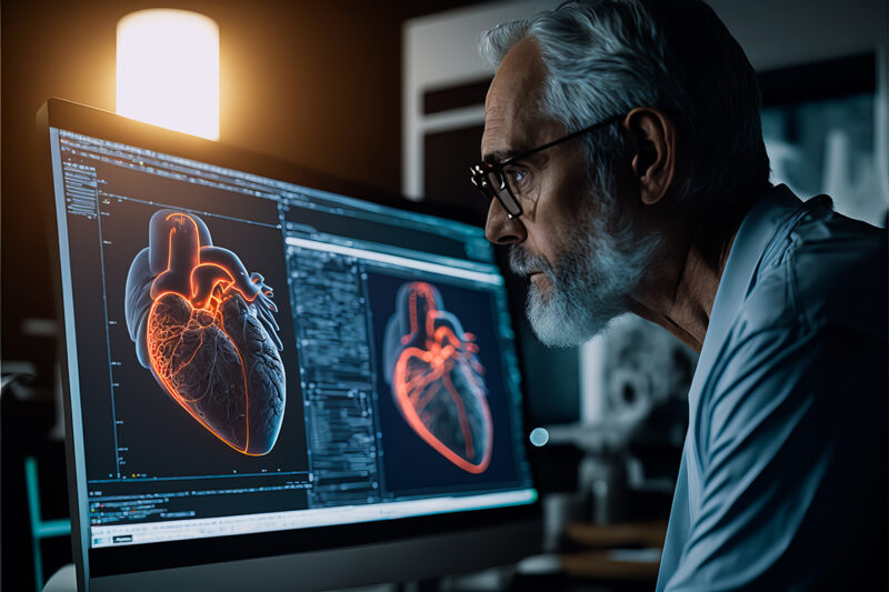
Recent years have witnessed several advancements in the field of cardiac imaging, giving radiologists access to higher-quality images for quicker and more precise interpretations. With modern medical imaging, physicians can now see in three dimensions and get images of the circulatory system, including the heart, that were previously unattainable. Thanks to these developments, physicians can now diagnose and predict patients more accurately, helping them avoid heart failure and identifying and evaluating cardiac issues much sooner.
Imaging using Magnetic Resonance (MRI)
Recent years have seen amazing breakthroughs in magnetic resonance imaging, allowing for a thorough evaluation of the anatomy and function of the heart. Specialised cardiac coils and high-field strength magnets have greatly improved image resolution.
Furthermore, methods like cine MRI provide real-time visualisation of the heart’s motion, which helps identify anomalies. When it comes to the diagnosis of illnesses of the congenital heart, myocardial infarction, and cardiomyopathies, magnetic resonance imaging is very helpful. MMI uses modern MRI technology, a specialist cardiologist dubai, to provide precise diagnosis and direct treatment plans.
Angiography using Computed Tomography (CT)
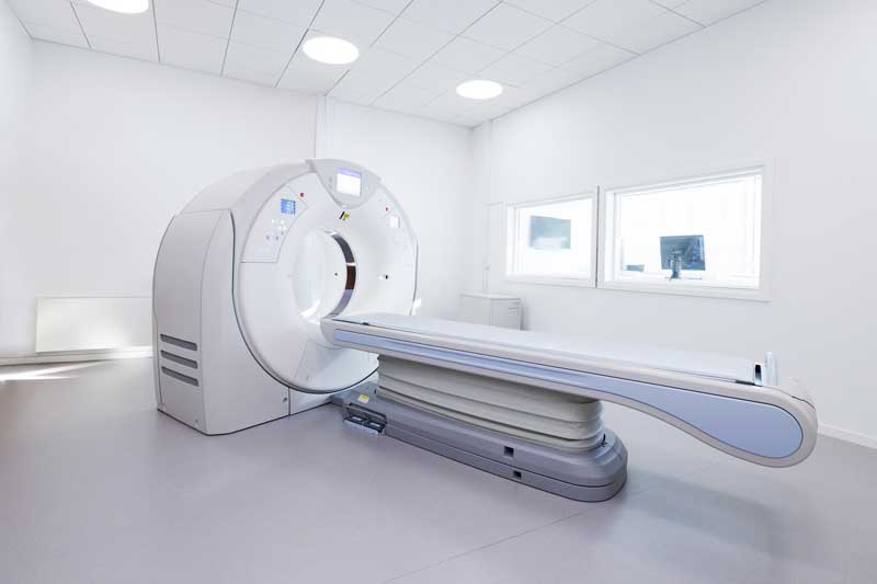
One effective method for visualising the cardiovascular system is CT angiography. It includes creating intricate cross-sectional pictures of blood arteries using computer algorithms and X-rays. Recent developments in CT technology allow for the acquisition of better-quality pictures at reduced radiation doses, guaranteeing patient safety. CT angiography is very helpful when assessing aortic illnesses, pulmonary embolisms, and coronary artery disease. CT angioplasty is often used at a cardiology facility in Dubai to determine the extent of arterial obstructions and to schedule procedures appropriately.
Echocardiography
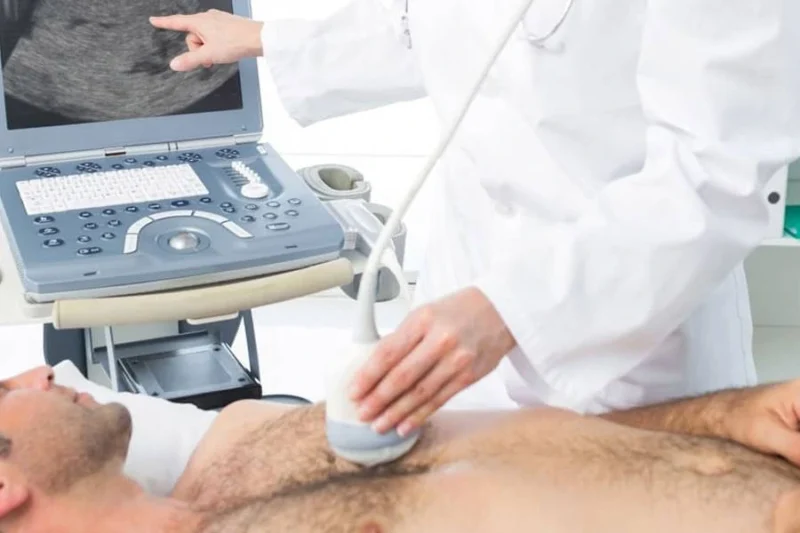
Heart ultrasound, or echocardiography, has long been a staple of cardiovascular imaging. The advent of sophisticated methods like strain imaging and three-dimensional echocardiography results from recent advancements. Comprehensive volumetric data on the cardiac chambers, controls, and genetic defects may be obtained by 3D echocardiography. By measuring the deformity of cardiac tissue, strain imaging can identify early indicators of heart illness and provide important information about how the heart works. The diagnosis of diseases such as congenital heart abnormalities, heart failure, and valvular heart disease is greatly aided by these developments in echocardiography.
Positron Emission Tomography (PET) Imaging
Non-invasive evaluation of cardiac metabolism and perfusion is now possible thanks to PET imaging, which has completely changed the area of molecular imaging. The accurate anatomical identification of functional abnormalities is made possible by the combination of PET and computed tomography, or PET/CT. When determining the degree of myocardial infarction, detecting regions of ischemia, and evaluating myocardial viability, PET imaging is very helpful. PET imaging is used by MMI Cardio Hospital in Dubai to determine the metabolic activity of heart tissues precisely and to schedule the necessary procedures.
Cardiac Angiography and Catheterization
Even though they are not new methods, heart catheterisation and radiography have advanced significantly in the last several years. The discipline has changed due to the advent of cutting-edge imaging technologies, including optical coherence tomography, also known as OCT, and intravascular ultrasonography (IVUS). IVUS allows doctors to see within the coronary arteries, calculate the amount of plaque present, and precisely plan procedures. OCT provides high-resolution coronary artery wall imaging, which helps identify susceptible plaques and evaluate stent placement. These cutting-edge imaging methods are used during cardiac catheterisation treatments in Dubai’s cardiac facilities to guarantee the best possible results.
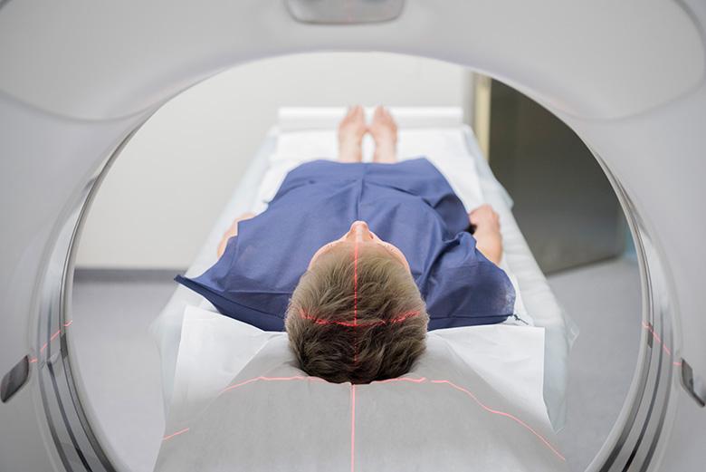
Cardiac Nuclear Imaging
Advanced nuclear imaging for coronary heart disease produces accurate, high-quality pictures of the circulatory system and blood flow. A tiny amount of radioactive tracer is injected into your bloodstream during this test, and it travels through the region that is being examined. A camera detects the tracer’s gamma rays and uses the information to map blood flow, assess cardiac muscle function, and evaluate coronary arteries. The test will take place in a medical lab, and your physician may order a computerised tomography (CT), positron emission tomography (PET), or magnetic resonance imaging (MRI) scan, depending on the kind of image they want to obtain.
These scans can produce unique views specific to your heart’s circumstances. Using these tests, our cardiologists can identify significant cardiac conditions and obstructive coronary circulation blockages. These conditions may necessitate angioplasty, which is the surgical repair of damaged blood vessels, or open heart bypass surgery to return normal cardiac blood flow. With tens of thousands of nuclear imaging studies previously completed, our cardiologists and technologists are qualified to critically assess and provide professional guidance about a course of treatment for your particular cardiac issue.
In summary
Cardiologists can now reliably detect and treat a broad spectrum of cardiovascular disorders because of advancements in cardiovascular imaging technology, which have completely changed the discipline of cardiology. Significant progress has been made in all these techniques, making it possible to see the circulatory system and arteries in great detail. These technologies support accurate diagnosis, tailored treatment planning, and early abnormality identification. Advanced imaging tools are essential to the high-quality care that patients with cardiovascular diseases get at Cardiology Institute in Dubai.